About the home page images
The structures shown on the BSG home page, the techniques and UK facilities behind them, and full credits.
Each image on the home page shows one molecule studied with several techniques, all at UK national facilities: bio-SAXS on beamline B21 and X-ray crystallography at Diamond Light Source, and cryo-EM at the Electron Bio-Imaging Centre (eBIC). A different image is shown each day.
The images were prepared for the BSG from public data (SASBDB, EMDB and PDB) and the published papers. We thank the authors for making their data openly available.
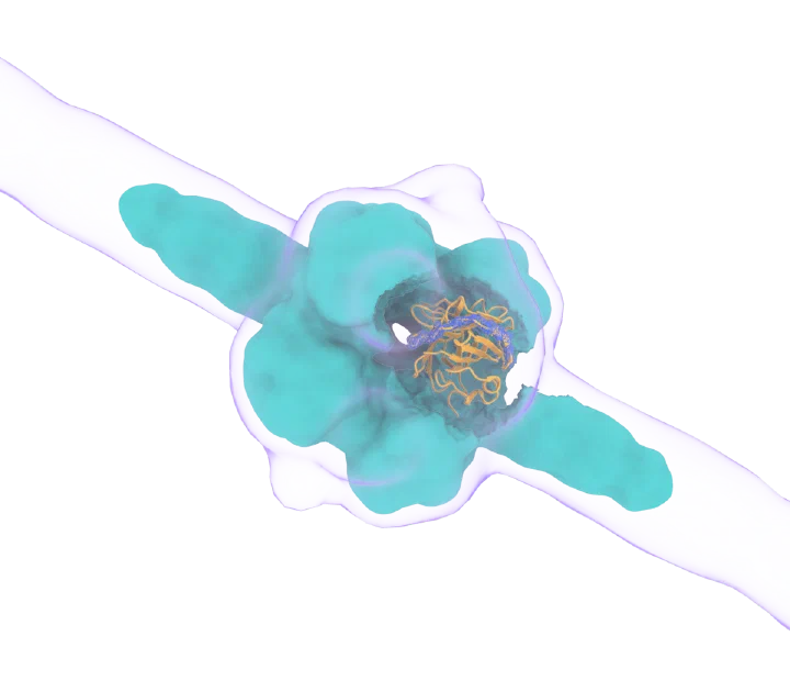
Pentraxin-3 (PTX3)
An innate-immunity protein that helps build the extracellular matrix, seen at three scales.
Tony Day's group, University of Manchester
Bio-SAXS · Diamond B21
Whole protein; the arms are an AlphaFold model validated against the SAXS data
Cryo-EM · eBIC
Central core, 3.3 Å
X-ray crystallography · Diamond I04-1
One domain at 2.4 Å, with electron density (blue)
- Data:
- SASBDB SASDVA6 · EMD-19717 · PDB 8S50 · PDB 8PVQ
- How it was made:
- The SAXS envelope is a low-resolution map of the authors' SAXS-validated model (SASDVA6). The X-ray density is the PDBe 2Fo−Fc map. Rendered with UCSF ChimeraX.
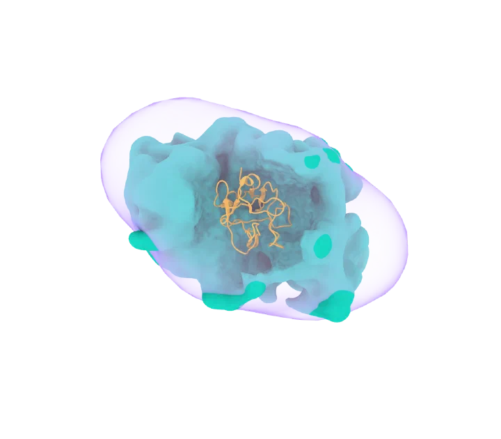
Phospholipase A2 receptor (PLA2R)
The kidney protein targeted by autoantibodies in membranous nephropathy; the window shows the domain carrying the dominant epitope.
Rachel Lennon's group, University of Manchester
Bio-SAXS · Diamond B21
Compact form at pH 6.2
Cryo-EM · eBIC
3.4 Å
Atomic model · from the cryo-EM map
Cysteine-rich domain
- Data:
- SASBDB SASDNQ3 · EMD-14077 · PDB 7QSR
- How it was made:
- The SAXS envelope was computed from the SASBDB data with DENSS (Grant, Nature Methods 2018). Rendered with UCSF ChimeraX.
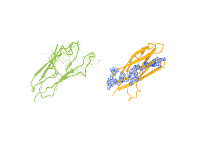
A new protein fold in the centrosome
Two domains of the centrosome scaffold protein Spd-2, one solved by NMR and one by crystallography, share a newly described fold.
Jordan Raff's group, University of Oxford, with Susan Lea (NCI, USA, and Oxford), Mark van Breugel (MRC LMB) and Zhe Feng (Fudan University)
NMR · Bruker 500/600 MHz
ASH1 domain, ensemble of models
X-ray crystallography · Diamond I04-1
ASH3 domain, 1.93 Å, with electron density (blue)
- Data:
- PDB 9FU8 · BMRB 34925 · PDB 9C72
- How it was made:
- The two domains were oriented the same way by structural alignment (TM-align; TM-score about 0.5, the same fold). The paper does not name the NMR facility. Rendered with UCSF ChimeraX.
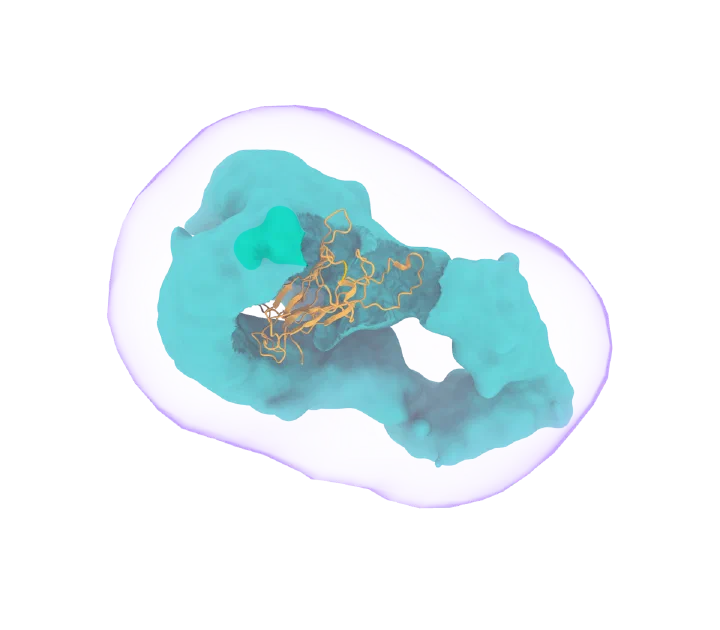
Toll5A receptor with Spätzle
A mosquito immune receptor pair bound to its signal protein, from the insect that carries dengue and Zika.
Monique Gangloff's group, University of Cambridge
Bio-SAXS · Diamond B21
The complex in solution
Cryo-EM · eBIC
4.2 Å
Atomic model · from the cryo-EM map
Spätzle ligand
- Data:
- SASBDB SASDKY8 · EMD-11982 · PDB 7B1B
- How it was made:
- The SAXS envelope was computed from the SASBDB data with DENSS (Grant, Nature Methods 2018). Rendered with UCSF ChimeraX.
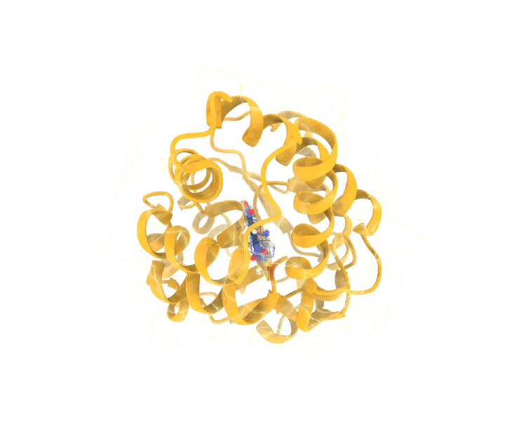
An enzyme evolved for pharmaceutical chemistry
A designed enzyme, improved by directed evolution, that performs a key pharmaceutical reaction with precise control of the product's handedness.
Anthony Green's group, Manchester Institute of Biotechnology
X-ray crystallography · Diamond I03
1.7 Å, substrate in electron density (blue)
Molecular dynamics and docking · GROMACS, AutoDock Vina
Showed how Arg124 holds the substrate in a reactive position
- How it was made:
- Crystal structure with the 2Fo−Fc density (PDBe) around the bound substrate. The enzyme was improved by directed evolution and characterised by kinetics and protein mass spectrometry; docking (AutoDock Vina) and molecular dynamics (GROMACS 2018, AMBER14 force field) were performed at the Manchester Institute of Biotechnology. The simulations are described in the paper, not shown in the image. Rendered with UCSF ChimeraX.
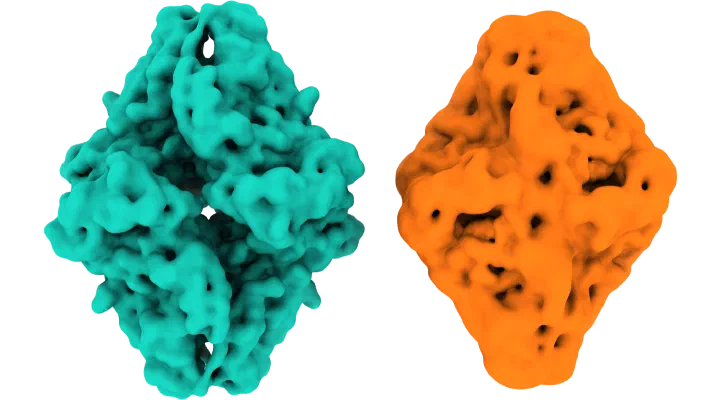
A protein that flew through a mass spectrometer
The enzyme β-galactosidase keeps its structure after passing through a mass spectrometer, answering a long-standing question for native mass spectrometry.
Stephan Rauschenbach's and Carol Robinson's groups, University of Oxford
Native mass spectrometry + cryo-EM · Orbitrap UHMR, soft landing
Mass-selected in the gas phase, then imaged, 2.6 Å
Cryo-EM · from solution
Conventional sample (Bartesaghi et al., Structure 2018)
- How it was made:
- Both maps filtered identically (Gaussian, 2 Å) and drawn to enclose the same volume, after aligning them by map-to-map fitting (correlation 0.90). The gas-phase structure is slightly more compact, as the paper reports. Rendered with UCSF ChimeraX.
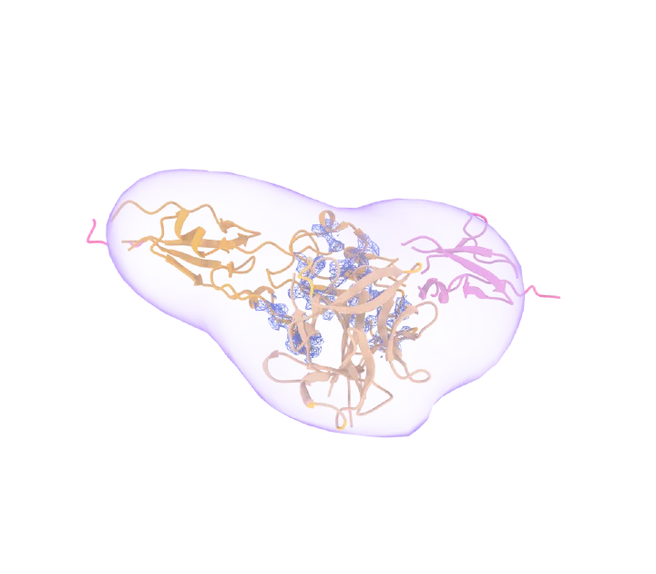
IL-33 held by a parasite protein
A protein from a parasitic worm that blocks IL-33, an alarm signal in allergy and asthma, seen by crystallography, SAXS and AlphaFold together.
Matthew Higgins's group, University of Oxford
Bio-SAXS · Diamond B21
The complex in solution
X-ray crystallography · Diamond I04
2.1 Å, with electron density (blue)
AlphaFold · validated by SAXS
Extra domain not seen in the crystal
- Data:
- SASBDB SASDUY6 · PDB 8Q5R
- How it was made:
- The SAXS envelope was computed from the SASBDB data with DENSS (Grant, Nature Methods 2018) and agrees with the authors' AlphaFold model deposited with SASDUY6 (correlation 0.93). The X-ray density is the PDBe 2Fo−Fc map. Rendered with UCSF ChimeraX.
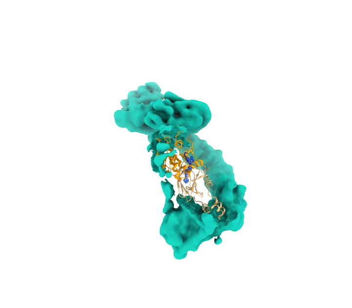
A degrader drug for cancer-causing KRAS
A small molecule that glues cancer-causing KRAS to the cell's disposal machinery, degrading 13 of the 17 most common KRAS mutants.
Alessio Ciulli's group, University of Dundee, with Boehringer Ingelheim
X-ray crystallography · SLS
KRAS–degrader–VHL complex, 2.2 Å, with electron density (blue)
Cryo-EM · Titan Krios
Full ligase complex, 3.5 Å
- How it was made:
- The crystal structure was superposed on the cryo-EM model through VHL; the cryo-EM surface is cut away around it. The X-ray density is the PDBe 2Fo−Fc map. Rendered with UCSF ChimeraX.